Successfully observed the state of cells inside cancer tissue models without labeling or destroying them
がん組織モデル内部の細胞状態を "無標識・非破壊”で観察することに成功
A joint research group led by Kazuyo Ito, Assistant Professor of Tokyo University of Agriculture and Technology Graduate School Institute of Engineering Division of Advanced Applied Physics, Yuta Iijima, Graduate School of Bio-Applications and Systems Engineering Department of Food and Energy Systems Science (4th year of 5-year integrated doctoral program), Tomoki Misumi, Graduate School of Engineering Department of Industrial echnology and Innovation (2nd year master's program), Professor Kenji Ikushima, Institute of Engineering Division of Advanced Applied Physics, Daisuke Yoshino, Associate Professor, Gen Hayase, a Unit Chief at the National Institute for Materials Science (NIMS), and Kazuki Tamura, Assistant Professor at the National Institute of Photonics, Hamamatsu University School of Medicine, we have succeeded in visualizing the three-dimensional structure without labeling and non-destructively.
The results of this research were published in npj Biomedical Innovations (June 13th).
Paper title: Biochemical state in tissue can be detected through ultrasound signal
URL:
https://www.nature.com/articles/s44385-025-00026-w
Previous research related to this paper
Paper title: Biological characterization of breast cancer spheroid formed by fast fabrication method
Published in: In vitro models, Volume 3, pages 19–32, 2024.
URL: https://doi.org/10.1007/s44164-024-00066-3
Press release: "Easily-prepared cancer tissue models open new possibilities in cancer research"
URL: https://www.tuat.ac.jp/outline/disclosure/pressrelease/2023/20240214_01.html
background
Spheroids are cell aggregates in which cells are aggregated into a spherical shape, and are characterized by three-dimensional culture rather than conventional two-dimensional culture on cell culture dishes. By culturing in three dimensions, we have developed a cancer tissue model (cancer spheroids) that reproduces an environment close to our body. Note 1) is becoming increasingly important in cancer research and drug discovery. However, it has been a long-standing task to observe changes in the cellular state inside cancer spheroids without destroying or dyeing (without labeling). Conventional optical microscopes have limited depth of observation, making it difficult to capture the inside of thick cancer spheroids in detail, which had to be overcome.
Research Structure
This research was supported by the Nakatani Medical Engineering Measurement Technology Foundation (Grant-in-Aid for Encouragement), JSPS Grant-in-Aid for Scientific Research (21K19893), and Terumo Life Science Foundation, and was conducted at Tokyo University of Agriculture and Technology, the National Institute for Materials Science (NIMS), and Hamamatsu University School of Medicine.
Research results
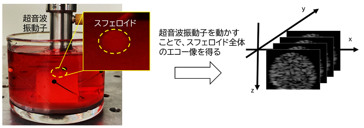
In this study, we applied ultrasound observation (Note 2), which is widely used in medical settings as an echocardiogram, to successfully observe the internal structure of cancer spheroids in their original form (Figure 1). This made it possible to "peek" at important changes such as internal shrinkage and cell death in real time and non-destructively.
First, based on the dynamic changes inside cancer spheroids revealed by the research group in a previous study (Iijima et al.,
In vitro models
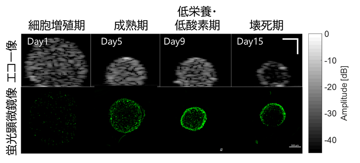
2024), particularly contraction, programmed cell death (apoptosis), and cell necrosis, the researchers clarified how these changes are reflected in ultrasound signals. They analyzed the spatial changes in echogenicity (Note 3) and brightness density inside the cancer spheroids and identified brightness patterns associated with cell proliferation, apoptosis, and necrosis (Figure 2). In particular, they confirmed that the brightness in the center of the cancer spheroids rapidly decreased after the third day of culture, and tended to be almost zero by the 15th day, suggesting the formation of a necrotic core (Note 4).
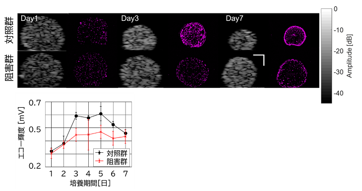
Furthermore, when the activity of actomyosin, a type of cytoskeleton involved in spheroid contraction, was inhibited with a drug, a significant change in the brightness pattern occurred, indicating that cell mechanics plays an important role in the formation of the internal structure of spheroids (Figure 3).
These results demonstrate that ultrasound, taking advantage of its characteristics of "deep reach," "no labeling required," and "real-time observation," is a promising new analytical method for not only cancer spheroids, but also 3D culture systems such as spheroids in general and organoids (Note 5).
Future developments
This study demonstrated that ultrasound can be used to visualize dynamic changes inside cancer spheroids nondestructively and without labels. In the future, it is expected that the spatial resolution of this method will be improved and applied to drug response evaluation and malignancy assessment of tumors, and that the application of this method will be expanded to co-culture models and organoids that are closer to living organisms, thereby contributing to the development of cancer research and regenerative medicine.
Glossary
Note 1) Cancer spheroids
A large number of cancer cells gather and aggregate to form a spherical mass.
Note 2) Ultrasound observation
A technology that uses high-frequency sound (ultrasound) that is too high for our ears to hear, to observe the inside of the body and the state of cells. Known as "echo examination" in the medical field, it is widely used for observing fetuses and diagnosing internal organs. It is characterized by being a safe observation method that is non-destructive and does not use radiation.
Note 3) Echo brightness
An index showing how bright each part appears in an image obtained using ultrasound. This brightness is determined by the strength of the ultrasound waves that hit the inside of cells or tissues and return to the ultrasound probe. Areas with high echogenicity indicate that the ultrasound waves are returning strongly, suggesting that cells are densely packed together or that there are structures with different acoustic properties.
Note 4) Necrotic core
A mass of necrotic cells inside a spheroid. Also called a necrotic core.
Note 5) Organoid
Artificially engineered three-dimensional cell aggregates that mimic biological organs and tissues.
◆Inquiries about research◆
Tokyo University of Agriculture and Technology Graduate School Institute of Engineering
Division of Advanced Applied Physics Assistant Professor
Kazuyo Ito
TEL/FAX:042-388-7807
E-mail: itokazuyo (please put @ here)go.tuat.ac.jp
◆ Inquiries about the press ◆
Tokyo University of Agriculture and Technology, General Affairs Office Public Relations Office
TEL:042-367-5930
E-mail: koho2 (please put @ here)cc.tuat.ac.jp
Hamamatsu University School of Medicine, General Affairs Office Public Relations Office Public Relations & Endowment Section
TEL:053-435-2151
E-mail: koho (please put @ here)hama-med.ac.jp
Related links (opens in a new window)
- Tokyo University of Agriculture and Technology Assistant Professor Kazuyo Ito Researcher Profile
- Tokyo University of Agriculture and Technology and Technology Professor Kenji Ikushima Researcher Profile
- Tokyo University of Agriculture and Technology and Technology Associate Professor Daisuke Yoshino Researcher Profile
- Tokyo University of Agriculture and Technology Daisuke Yoshino Associate Professor, Kazuyo Ito Assistant Professor Laboratory Website
- Tokyo University of Agriculture and Technology and Technology Professor Kenji Ikushima Laboratory Website
- Professor Kenji Ikushima, Daisuke Yoshino Associate Professor, and Kazuyo Ito Assistant Professor belong to Tokyo University of Agriculture and Technology Faculty of Engineering Department of Biomedical Engineering