Developed a three-dimensional cultured vascular model that replicates the forces acting on blood vessels – Enabling stent placement and contributing to next-generation stent design.
Development of a three-dimensional cultured blood vessel model that replicates the forces acting on blood vessels
– Enables stent placement and contributes to the design of next-generation stents –
Dr. Taku Okuno (at the time of research), Professor Daisuke Yoshino of the Graduate School of Graduate School of Engineering Department of Biomedical Engineering Tokyo University of Agriculture and Technology Institute of Global Innovation Research, and Assistant Professor Kazuyo Ito of the Graduate School of Institute of Engineering Division of Advanced Applied Physics, through joint research with Professor Kenichi Funamoto of the Institute of Fluid Science, Tohoku University, We have developed a three-dimensional culture vascular model that can actually place a stent, and succeeded in quantitatively evaluating the endothelialization process after stent placement under a physiological blood flow environment. In this study, we systematically constructed a series of experimental systems from the design and production of vascular models to cell culture, blood flow loading, stent placement, and evaluation methods, and the detailed protocols are specified in the paper. This made it possible to comprehensively analyze the dynamic effects of stents on the vessel wall and the dynamics of vascular endothelial cells in response to them under the same conditions.
This result is expected to deepen our understanding of the mechanism of restenosis and thrombosis, as well as contribute to the optimization of next-generation stent design.
The results of this research were published in BMC Methods (May 12th).
Paper title: Cell-cultured PDMS vascular model to allow placement of implant devices
URL: https://doi.org/10.1186/s44330-026-00072-9
Preprints related to this paper
Preprint URL: https://doi.org/10.1101/2025.01.20.634010
background
Stent placement is a widely used treatment for vascular stenosis caused by arteriosclerosis and other conditions. However, if excessive mechanical stress is applied to the vessel wall due to the difference in stiffness between the stent and the vessel wall, it can cause inflammation, thrombus formation, and restenosis (Note 1). For the long-term success of stent treatment, the progression of "endothelialization (Note 3)," in which vascular endothelial cells (Note 2) rapidly cover the stent surface after placement, is crucial. To understand this interaction between stents and blood vessels, an experimental system is needed that can reproduce and evaluate the vascular response after stent placement in a blood flow environment. However, until now,
- There were limited vascular models available in which stents could actually be placed.
- The force generated by blood flow could not be adequately reproduced.
- An experimental system for comprehensively quantitatively evaluating the mechanical environment and cellular responses had not yet been established.
These were some of the challenges we faced.
Research Structure
This research was supported by the Uehara Memorial Life Science Foundation (FY2020 Research Grant) and was conducted at Tokyo University of Agriculture and Technology and Tohoku University.
Research results
In this study, we developed a three-dimensional vascular model made of polydimethylsiloxane (PDMS) and formed a cylindrical endothelial monolayer by seeding human carotid artery-derived vascular endothelial cells into its lumen.
When cells were cultured in a perfusion environment that replicated blood flow by connecting a perfusion device, the endothelial cells elongated and oriented along the direction of flow, showing a response similar to that in vivo (Figure 1). Furthermore, it was revealed that the rate of cell loss changed depending on the blood flow conditions, demonstrating that the effect of forces acting on blood vessels can be quantitatively evaluated.
Furthermore, a self-expanding stent made of nickel-titanium alloy was placed in the model and cultured in a blood flow environment (Figure 2). As a result, approximately 20% of the stent's sides were covered with vascular endothelial cells after 24 hours and approximately 50% after 48 hours, indicating endothelialization (Figure 3).
This study is characterized by presenting an experimental system that allows for the three-dimensional and quantitative analysis of the processes of endothelial cell migration, adhesion, and coverage after stent placement. Furthermore, by specifically describing the apparatus configuration and experimental conditions, it proposes a practical method for evaluating the mechanical interaction (Note 4) between the stent and the vascular wall.
Future developments
In the future, we will further refine our approach by developing multilayered models that include vascular smooth muscle cells and by introducing materials with elastic properties closer to those of living organisms, thereby more precisely reproducing the mechanical environment of the blood vessel wall. Furthermore, by combining this with visualization techniques for the stress distribution between the stent and the blood vessel wall, we plan to develop an evaluation system that contributes to elucidating the mechanisms of restenosis and optimizing stent design.
Glossary
Note 1) Restenosis
This phenomenon involves the narrowing of blood vessels that have been dilated by treatment. Excessive cell proliferation and inflammation in the blood vessel walls are involved.
Note 2) Vascular endothelial cells
These cells make up the innermost layer of cells that line blood vessels. They are directly exposed to blood flow and play a crucial role in maintaining vascular function.
Note 3) Endothelialization
This phenomenon involves vascular endothelial cells covering the stent surface after stent placement. Sufficient endothelialization helps suppress thrombus formation and restenosis.
Note 4) Mechanical interaction
The relationship between the mechanical action between the stent and the blood vessel wall, and the blood vessel's response to that action.
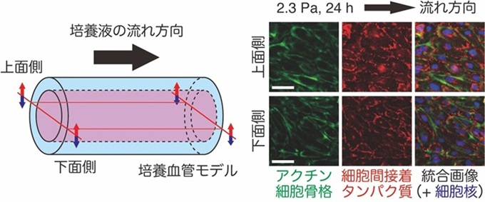
Figure 1: Immunofluorescence staining image of vascular endothelial cells covering the inside of a cultured blood vessel model. The blood vessel model was subjected to a shear stress of 2.3 Pa for 24 hours, then cut in half vertically, stained, and observed. In response to the flow of culture medium simulating blood flow, the vascular endothelial cells can be seen aligning and extending in the direction of flow. The scale bar represents 50 µm. The figure was created by Yoshino et al., modified from (Okuno et al, 2026. BMC Methods, 3, 16).
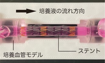
Figure 2: A self-expanding stent made of nickel-titanium alloy is placed in the developed cultured blood vessel model. The figure was created by Yoshino et al., modified from (Okuno et al, 2026. BMC Methods, 3, 16).
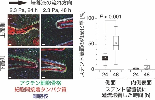
Figure 3: Stent endothelialization. You can observe how vascular endothelial cells on the inner surface of a cultured blood vessel model proliferate and cover the sides of the stent (the area enclosed by the white wavy line shows the part of the stent surface covered by vascular endothelial cells). The scale bar represents 100 µm. The figure was created by Yoshino et al., modified from (Okuno et al, 2026. BMC Methods, 3, 16).
◆Inquiries about research◆
Tokyo University of Agriculture and Technology Institute of Global Innovation Research
Professor Daisuke Yoshino
TEL/FAX:042-388-7113
E-mail: dyoshino (please put @ here)go.tuat.ac.jp
◆Inquiries about the press◆
Tokyo University of Agriculture and Technology Public Relations Office
E-mail: koho2 (put @ here)cc.tuat.ac.jp
Institute of Fluid Science, Tohoku University International Research Strategy Office (Public Relations)
E-mail: ifs-koho (put @ here)grp.tohoku.ac.jp
Related links (opens in a new window)
- Tokyo University of Agriculture and Technology Professor Daisuke Yoshino Researcher Profile
- Tokyo University of Agriculture and Technology Assistant Professor Kazuyo Ito Researcher Profile
- Tokyo University of Agriculture and Technology Prof. Daisuke Yoshino and Asst. Prof. Kazuyo Ito Laboratory website
- Professor Daisuke Yoshino and Assistant Professor Kazuyo Ito belong to Tokyo University of Agriculture and Technology Faculty of Engineering Department of Biomedical Engineering