Mast cell hyperactivity underpins the development of oxygen-induced retinopathy
April 2, 2018
Professor Hiroshi Matsuda (Division of Animal Life Science, Institute of Agriculture, Tokyo University of Agriculture and Technology) and his research group have clarified the complete mechanism of the pathogenesis of murine oxygen-induced retinopaty, a model for retinopathy of prematurity that has not been known so far. In addition, they detected a high level of the pathogenic factor in perpheral blood of the babies with retinopathy of prematurity. Based on these results, it is expected that the early diagnosis method of this disease and the novel prevention method and therapy will be developed in the future. This research was publised on October 10 in the Journal of Clinical Investigation.
The eye provides a window into the process of vascularization in both health and disease. Ocular neovascularization is the leading cause of blindness in adults, for instance in age-related macular degeneration and diabetic retinopathy. In infants and children, retinopathy of prematurity (ROP) is also a leading cause of blindness, induced by oxygen-dependent retinal vascular injury and occlusion, and in severe cases subsequent neovascularization with a risk of retinal scarring and detachment. ROP only develops in preterm infants, particularly those at the extremes of survival and thus provides an insight at the interface between normal and abnormal vascular development. Excessive oxygen therapy given to extremely preterm infants is the key risk factor for ROP. It results in vascular occlusion and subsequent reactive neovascularization mediated by VEGF, which can be partly offset using anti-VEGF monoclonal therapy. However, some ROP patients still end up with visual impairment, even after treatment. The precise mechanism by which aberrations in infant’s oxygen tension triggers neovascularization is currently unclear.
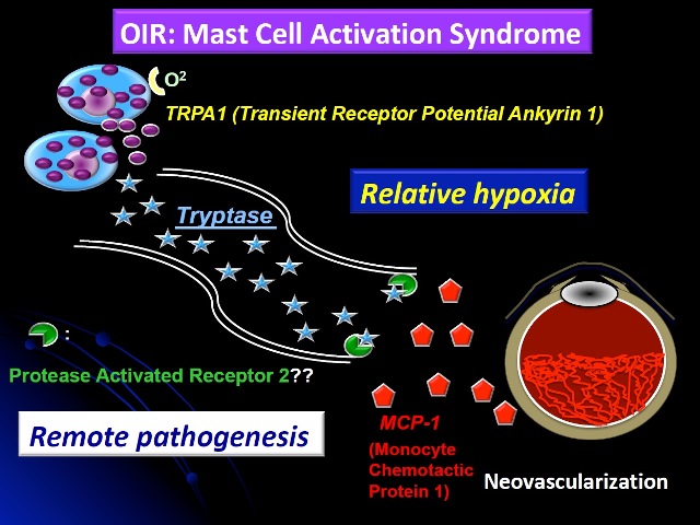
Although mast cells are classically thought of as protecting the host against helminth infections and inducing allergic diseases, recent studies have revealed their additional role in neovascularization, which is critical for tissue remodeling, chronic inflammation, and carcinogenesis. Here we demonstrate that mast cells are essential for sprouting angiogenesis in oxygen-induced retinopathy (OIR). Mouse strains lacking mast cells did not develop retinal neovascularization. OIR could be induced in these mice by infusion of mast cells or by injection of mast cell tryptase (MCT). Relative hypoxia stimulated degranulation of mast cells via transient receptor potential ankyrin 1. Subsequent surges in MCT induced the production of monocyte chemotactic protein-1 (MCP-1) and angiogenic factors by retinal endothelial cells leading to sprouting angiogenesis. Mast cell stabilizers, as well as specific tryptase and MCP-1 inhibitors prevented the development of OIR in wild type mice (Fig. 1). To demonstrate the relevance of these findings in humans, we also showed that preterm infants developing retinopathy of prematurity have significantly higher plasma MCT levels than age-matched controls.
Our results suggest that therapies which suppress mast cell activity may have the potential for preventing eye diseases and subsequent blindness induced by neovascularization.
Paper
| Authors | K. Matsuda, N. Okamoto, M. Kondo, P.D. Arkwright, K. Karasawa, S. Ishizaka, S. Yokota, A. Matsuda, K. Jung, K. Oida, Y. Amagai, H. Jang, E. Noda, R. Kakinuma, K. Yasui, U. Kaku, Y. Mori, N. Onai, T. Ohteki, A. Tanaka, and H. Matsuda |
|---|---|
| Title | Mast cell hyperactivity underpins the development of oxygen-induced retinopathy |
| Journal | Journal of Clinical Investigation |
| DOI | 10.1172/JCI89893 |
| URL | https://www.jci.org/ |
Acknowledgment
This work was supported by Grant-in-Aid for Scientific Research grants S #16H06383 (to H. Matsuda) and A #15H02478 (to A. Tanaka), provided by the Japan Society for the Promotion of Science.
Related Information
TUAT research factors (English): http://www.rd.tuat.ac.jp/en/activities/factors/index.html
Researcher information (English):Hiroshi Matsuda
Press release (Japanese): 未熟児網膜症の発病機構の全容を疾患モデルマウスを用いて解明 (PDF:361KB)
Contact
Hiroshi Matsuda
Professor, Division of Animal Life Science, Institute of Agriculture, Tokyo University of Agriculture and Technology
Tel +81-42-367-5784